SBI3U - Mr. SMITH BIOLOGY
Welcome to your interactive science experience. You will be able to view lesson materials, copy worksheets, and get review work from this interactive website. This website will have links posted for you to follow to gain a more in-depth understanding of the topics that we cover in class. Now, scroll thorugh, click on the links, send me emails and enjoy! (pg ref. from BIOLOGY 11 McGraw-Hill Ryerson textbook)
Saturday, May 14, 2005
HUMAN DIGESTIVE SYSTEM SUMMARY
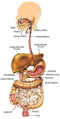
Mammalian Digestive System:
This is a basic overview of what occurs during the 4 stages of attaining nutrients for the body.
This overview will indicate the various pathway that your nutrients will follow. Ingestion, Digestion, Absorbtionand finally to Excretion
Oral cavity: Both physical and chemical digestion takes place in the mouth.
Saliva is secreted to moisten food, protect the mouth from abrassions, buffer against acids in food, kill some forms of bacteria, and begin carbohydrate digestion with the enzyme SALIVARY AMYLASE.
TONGUE: is used for taste, manipulates food while chewing and prepares food for swallowing by forming it into a ball.
Pharynx: Commonly called the throat. The intersection of the glottis and opening to the esophagus (gullet) is found here. The epiglottis is a flap that closes the glottis when the act of swalloing occurs.
Esophagus: Connects the pharynx and the stomach. Peristalsis, wave-like contractions of the smooth muscles push food down toward the stomach. Connects with the stomach at the CARDIAC SPHINCTER.
Stomach: J shaped expandable organ located on the left side of the abdominal cavity.
Stores up to 2 liters of food while mixing and digesting it. The epithelial cells secrete GASTRIC JUICES and HCl making the pH around 2.
PEPSIN is an enzyme used to partially hydrolyze protein. Pepsin is released in an inactive form PEPSINOGEN. The pepsinogen reacts with HCL to form pepsin.
The hormone GASTRIN is secreted by the stomach cells to regulate the production of gastric juices. The stomach is closed at its posterior end by the PYLORIC SPHINCTER.
Small Intestine: Most hydrolysis of macromolecules occur in the small intestine. It is more than 6 meters in length. It has smaller diameter than that of the large intestine.
It is divided into 3 sections ( Duodenum, Jejunum, and ileum). Accessory Organs ( Pancreas, Liver, and Gall Bladder), add digestive enzymes, juices and hormones into the small intestine. As the acid chyme enters the duodenum (first 25 cm of the small intestine) a hormone called SECRETIN is released from the intestinal walls to siginal the pancreas to release a bicarbonate solution which neutralizes the acid.
The hormone CHOLECYSTOKININ (CCK), is released from the intestinal cells causing the gall bladder to release bile. It also causes the pancreas to release its digestive enzymes. The hormone ENTEROGESTRONE is also secreted to slow down peristalsis. Protein Digestion: Trypsin and Chymotrypsin are enzymes that break bonds next to specific amino acids. Carboxypeptidase splits off one amino acid at a time.
This enzyme works on the end with the free carboxyl group. Aminopeptidase works in the opposite direction. All the above enzymes are secreted in an inactive form. They are activated by the hormone ENTEROKINASE. Fat Digestion: Bile emulsifies fat. This creates a larger surface area for the enzyme lipase to digest it. Carbohydrate Digestion: Disaccharide digestion is under the control of the enzymes maltase, lactase, sucrase.
ABSORPTION AND DISTRIBUTION OF NUTRIENTS:
The lining of the small intestine has a surface area of 600m2 , about the size of a baseball diamond. Large folds are decorated with fingerlike projections called villi, and each of the epithelial cells has many microscopic appendages called microvilli. The nutrients, except fat, are absorbed into the capillaries, while the fat enters the lacteal. Only 2 single layers of epithelial cells separate the lumen from the bodies blood supply. In the center of the villus is a tiny lymphatic vessel called the lacteal. Fatty substances enter the lacteal, along with other materials too large to enter the capillaries. This material is dumped into the blood neear the left shoulder ( thoracic duct ). Most of the nutrients are pumped against a concentration gradient by epithelial membranes. Sodium is pumped out of the cells into the lumen and fuels the entry of the nutrients into the cells and blood vessels by passively flowing back into the cells with the nutrients. All the contents of the blood enter the liver via the Hepatic portal vein. The liver regulates the contents of the blood.
Large Intestine: The large intestine is connected to the small intestine at a T-shaped junction where a blind pouch called the cecum is found. The cecum ends with a small fingerlike projection called the appendix. The function of the colon is to reabsorb water from the unused food. It reabsorbs 90% of the 7 liters of fluid secreted by the digestive tract. Solid waste is called feces. The bacterium E.coli produces odoriferous gases such as methane and hydrogen sulfide, while others produce vitamin K. The terminal portion of the colon is the rectum where the feces are stored until elimination from the body.


